Cornea Keratoconus
Cornea Cross-Linking
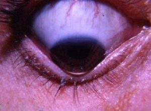 Keratoconus is an eye condition where the cornea thins and causes a distortion to its surface.
Nearly 90% of the cornea is made of overlaid collagen fibrils that are interconnected with cross-links to give the cornea strength and stability. When the cornea distorts in shape, patients complain of poor vision – notably, nighttime haloes and streaks.
There are a number of options to improve vision but none of them stops the disease from progressing. In some patients, this means that there is a need for a corneal transplant. The results are variable and patients still need to wear glasses or contact lenses. Furthermore, complete healing can take more than a year. In order to maintain the health of the graft, long-term eye drops are also required.
In order to reduce the likelihood of corneal transplant surgery, it is advisable to consider corneal cross-linking treatment. Cross-linking treatment aims to prevent the progression of keratoconus. However, it will not reverse the distortion.
Once the eye is numbed with drops, further drops of Riboflavin are added to the eye. Riboflavin is instilled until the cornea is saturated. A small dose of UV-A light is used to activate the Riboflavin. This increases the number of cross-links, resulting in a strengthened cornea. Cross linking is the sensible option for patients with mild to moderate progressive keratoconus.
ICRS Suitability
 The irregular shape of the cornea in keratoconus causes a distortion to vision, especially at night.
The intracorneal rings surgical procedure is designed to improve vision in patients with keratoconus where glasses and contact lenses are no longer tolerated. Intracorneal rings can also postpone the need for a corneal transplant. Once the rings are implanted, they are virtually invisible to the naked eye.
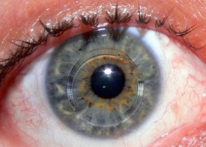
Topography
 Corneal topography is an advanced technology to measure the shape of the cornea. Modern-day topographers can measure thousands of points in the cornea, resulting in an ultra-high definition assessment of its profile. Corneal topography measurement is painless and takes less than a minute.
As the cornea is the most powerful optical component of the eye, any changes to its shape can adversely affect vision. Advanced computer calculations provide your practitioner with colour-coded maps that inform your practitioner about the presence of conditions like keratoconus, where the cornea becomes an irregular shape. Corneal topography is invaluable during complex contact lenses fitting to better align the lens with the shape of the eye. Furthermore, anyone having any form of laser eye surgery, such as LASIK or PRK, will be required to have a topography scan before and after the procedure.
Transplant
![]() A corneal transplant involves the removal of the damaged cornea and replacing it with a donor cornea. Modern-day corneal transplantation has evolved significantly. If only the top layers of the cornea are damaged, your surgeon will transplant only the upper layers. This results in lower rejection rates, improved stability, and better vision. Your surgeon can perform a procedure called DSEK, where this thin layer is removed and replaced with donor tissue. Unfortunately, in some cases, the entire cornea may need to be replaced with a full thickness corneal graft.
A corneal transplant involves the removal of the damaged cornea and replacing it with a donor cornea. Modern-day corneal transplantation has evolved significantly. If only the top layers of the cornea are damaged, your surgeon will transplant only the upper layers. This results in lower rejection rates, improved stability, and better vision. Your surgeon can perform a procedure called DSEK, where this thin layer is removed and replaced with donor tissue. Unfortunately, in some cases, the entire cornea may need to be replaced with a full thickness corneal graft.